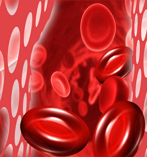
Spherocytosis
Hereditary Spherocytosis is a genetic disorder characterized by red blood cells that are fragile and spherical in shape instead of the normal flat disk shape. The abnormal shape makes it difficult for red blood cells to pass through the spleen. The spleen's function is to purify the blood and safeguard the immune function by fighting against the potentially dangerous bacteria, viruses or other microorganisms in the blood. In case of Spherocytosis, the membrane of the red blood cells is defective lending it a spherical shape. When these defective blood cells pass through the spleen, they break and destroy causing premature death of the red blood cells. The shortage of blood cells gives rise to severe anemia. It is an inherited type of Hemolytic Anemia. A parent with the disease has a 50% chance of having a child with the disease. Spherocytosis is most common in people of northern European descent.
Symptoms
The symptoms can vary from mild to severe. In severe cases, the disorder may be found in early childhood. In mild cases it may go unnoticed until adulthood. Patients with Spherocytosis exhibit all the symptoms associated with anemia such as fatigue, irritability, shortness of breath and muscle weakness. They also appear pale and weak. In addition to these symptoms, Spherocytosis also causes enlarged spleen and jaundice with yellow skin and eyes. Jaundice occurs due to the elevated levels of serum bilirubin as the red blood cells are destroyed within the body. Excess bilirubin, sometimes, also results in in the gallbladder. These stones may cause pain, infection or may block the tubes that lead out of the gallbladder.
Diagnosis
Family history is checked for hereditary factor and the abdomen has to be checked for enlarged spleen. Following blood tests may be performed to support the clinical examination:
- CBC or complete blood count test to check for anemia.
- The Reticulocyte Count Test is performed to measure the new and immature red blood cells produced in the bone marrow. The reticulocyte count is usually higher in cases of Spherocytosis.
- Osmotic Fragility Test is performed to measure the fragility of the red blood cells. This involves taking a blood sample and suspending in a salt solution and then measuring the destruction and fragility of the cells.
- Coombs Test to measure the antibodies in the blood.
- Blood test to check for bilirubin levels.
Treatment
Splenectomy is the most definitive treatment for hereditary Spherocytosis. By removing the spleen, red blood cells are prevented from damage and are allowed to stay alive for a longer duration. However, removing a spleen makes the patient susceptible to infections for a lifetime. Hence patients need to take penicillin (or another antibiotic) for the rest of their lives. Immunization against pneumococcal and meningococcal infections is also prescribed. In case of severe anemia, blood transfusion is advised.
Reticulocyte Count Test
Blood is a precious fluid. Plasma, white blood corpuscles and red blood corpuscles are the three kinds of cells in the blood. The blood cells are normally made in the bone marrow. To know the level of red blood cells in the blood, reticulocyte count test is done.
Reticulocyte Count Test
Reticulocyte are immature red blood cells made by the bone marrow. Reticulocytes are in the blood for two days before developing into mature cells. Reticulocyte count test is done to determine if red blood cells are being made in the bone marrow at an appropriate rate. The count indicates how quickly reticulocytes are being produced and released by the bone marrow.
Need for Reticulocyte count test
Paleness, tiredness, weakness, shortness of breath, and/or blood in the stool are symptoms when doctors recommend Reticulocyte count test to:
- Assess the functioning of bone marrow i.e. if enough red blood cells are made by the bone marrow.
- Diagnose and distinguish the different kinds of anemia and the reasons for anemia, if it is due to fewer red bloods or due to great loss of red blood cells.
- Evaluate reasons for chronic bleeding and or/blood in the stool.
- Monitor progress after chemotherapy, bone marrow transplant, treatment for iron deficiency etc.
- Determine the degree and rate of increased number of RBCs and elevated hemoglobin (rare occurrence) and hematocrit.
Reticulocyte count test - Preparation
There isn't any specific preparation required before Reticulocyte count test. However, it is recommended to check with health care provider if any specific pre test preparation is needed. Those who have had a recent blood transfusion should inform the doctor. This can affect test results. Important information to be shared with the doctor is about medicines being taken. Women who are pregnant and mothers who are breastfeeding infants should talk to doctor before opting for the test.
Reticulocyte count test Method
The test result is available few hours after collecting blood or the next day. An elastic band is wrapped around the upper arm to stop the flow of blood. Sample of blood from the vein of the arm is taken. A drop of blood is placed on a slide, smeared, stained and is examined under a microscope. This is the manual method. Automated methods that allow for greater number of cells to be counted are most likely to replace the manual method.
Reticulocyte count test result Interpretation
The number of reticulocytes is compared to the total number of red blood corpuscles (RBC) and is reported as a percentage of reticulocytes.
Reticulocyte (%) = [Number of Reticulocytes / Number of total Red Blood Cells] X 100
In a healthy adult person, the normal range is 0.5% to 1.5%. In kids, the normal range is 3% to 6%. This is a stable percentage. The normal range may slightly vary.
High reticulocyte count suggests more red blood cells are being made by the bone marrow. This could be due to acute bleeding, chronic blood loss, hemolytic anemia, kidney disease and potentially a fatal blood disorder in the fetus or new born - erythroblastosis fetalis (Erythroblastosis Fetalis refers to a serious blood disorder in infants - Rh incompatibility disease and ABO incompatibility disease) or hemolytic disease. Also, the count increases post treatment for certain anemia like iron deficiency anemia, pernicious anemia, or folic acid deficiency anemia.
Erythroblastosis Fetalis refers to a serious blood disorder in infants - Rh incompatibility disease and ABO incompatibility disease.
Low reticulocyte count suggests fewer red blood cells are being made by the bone marrow. This can be due to aplastic anemia or iron deficiency anemia. Other instances such as exposure to radiation, a chronic infection or due to intake of certain medicines (drug toxicity) can lead to low count.
The reticulocyte count increases temporarily during pregnancy. The count is high in newborns but within few weeks the count drops to adult levels. In case the result is abnormal more tests may be administered for further analysis.
Anemia
Anemia stands for 'without blood' in Greek; When the number of red blood cells (RBC) falls below normal, Anemia is a resultant condition. Hemoglobin is an important constituent of RBC. Hemoglobin usually occurs in the range of 12 and 18 g/dL (grams per deciliter of blood). If the hemoglobin levels show a decrease, anemic conditions set in. Consequently, the various organs and tissues of the body do not receive adequate oxygen on account of the diminished oxygen carrying capacity of the blood. This impairs their normal functioning. Usually women have smaller stores of iron than men. Besides, they also lose blood during menstruation making them primary targets for anemia.

World Health Organization (WHO) defines anemia as a hemoglobin level lower than 13 g/dL in men and lower than 12 g/dL in women. It is essential to be familiar with the typical symptoms of anemia. Often anemia is misdiagnosed and left untreated. An anemic person is likely to feel extremely tired and weak. This is accompanied with dizziness and breathlessness. A person suffering from anemia tends to appear pale and experience feelings of depression. In some cases, anemia can lead to heart ailments too.
Causes of Anemia
- Serious disease or infection such as hookworm infection, bleeding piles, esophageal var ices and peptic ulcers.
- Hemorrhagic - Excessive blood loss due to surgery, menstruation or injury.
- Genetic defects lead to sickle cell anemia, Thalassemia anemia and aplastic anemia.
- Hemolytic - Excessive intravascular blood destruction where red blood cells are destroyed prematurely.
Types of Anemia
Iron deficiency Anemia - Nearly 20% adult women tend to suffer from this form of anemia. Loss of blood due to menstruation is not compensated with an iron-rich diet. Pregnancy and breast feeding can also deplete iron stores. Iron deficiency anemia is also noticed during growth spurts or internal bleeding.
Aplastic anemia - When the bone marrow does not produce sufficient quantities of blood cells, aplastic anemia is noticed. Childhood cancers such as leukemia are often responsible for this form of anemia. Other possible causes of aplastic anemia are radiation, cancer or antiseizure medications and chronic diseases such as thyroid or kidney malfunction. Treatment for aplastic anemia involves blood transfusions and bone marrow transplant. This is done to replace malfunctioning cells with healthy ones.
Vitamin deficiency anemia - Low levels of folic acid lead to faulty absorption of iron. Anemia caused due to folic acid deficiency is called Megaloblastic anemia. Pregnancy doubles the body requirements of folic acid and it is imperative that pregnant women take folic acid supplements. Good dietary sources of folate are fresh fruits, green leafy vegetables, cruciferous vegetables, liver and kidney, dairy products and whole grain cereals. Vegetables should be eaten raw or lightly cooked.Folic acid anemia is also a common problem faced by alcoholics. Vitamin B-12 deficiency can lead to a condition of Pernicious anemia. Diseases such as thyroid malfunction or diabetes mellitus can affect the body's ability to absorb vitamin B-12. This vitamin is vital in the production of hemoglobin.
Vitamin C Deficiency Anemia is a rare form of Anemia that is the result of small red cells owing to prolonged dietary deficiency of the Vitamin C.
Sideroblastic Anemia: In this anemia, the body has sufficient iron but it fails to incorporate it into hemoglobin.
Hemolytic Anemia results from high rate of destruction of Red Blood Cells (RBC) at a rate faster than the rate bone marrow can replenish them.
Thalassemia anemia - Thalassemia or Cooleys Disease is a hereditary disorder found predominantly in people of South East Asian, Greek and Italian racial groups. This form of anemia is seen in differing degrees as Thalassemia encompasses a group of related disorders that affect the human body in similar ways. The most common occurrences of Thalassemia are alpha and beta thalassemia. Alpha thalassemia occurs when there are defects in the genes that produce alpha globin, while beta thalassemia occurs when there are defects in the genes that produce beta globin. The severity of the disorder depends on how many genes are affected and the specific mutations involved. Thalassemia anemia is characterized by symptoms like jaundice, enlarged spleen, shortness of breath and facial bone deformities.
Thalassemia is a group of inherited blood disorders that affect the production of hemoglobin, a protein in red blood cells that carries oxygen throughout the body. The disorder is caused by mutations in the genes that control the production of hemoglobin.
Thalassemia anemia occurs when a person has fewer red blood cells than normal, or the red blood cells are smaller and do not contain enough hemoglobin. This can lead to a range of symptoms, including fatigue, weakness, pale skin, jaundice, and an enlarged spleen.
Treatment for thalassemia anemia may involve blood transfusions, iron chelation therapy to remove excess iron from the body, and bone marrow transplant in severe cases. With appropriate treatment and management, many people with thalassemia can lead healthy and productive lives.
Diagnosing Anemia
A complete blood count test will test for hemoglobin levels and display an anemic condition. But often anemia is a symptom whose cause lies deeper. The cause and type of anemia will determine the treatment that is needed. A stool test will help in detecting occult blood. Hemoglobin electrophoresis is a blood test that helps identify abnormal hemoglobins. Diagnosing thalassemia or sickle cell anemia becomes possible with this test.
Treating Anemia
Deficiency can be treated with supplements of iron, Vitamin B-12 and Vitamin C. Partaking an iron-rich diet can be beneficial for those suffering from nutritional deficiency anemia. Seafood, nuts, whole grains and dried fruits such as raisins, prunes and apricots are rich in iron. Ensure adequate consumption of Vitamin C as it aids and stimulates iron absorption. Try and combine citrus foods with iron-rich foods - add tomatoes to a turkey sandwich or chopped strawberries with iron-fortified breakfast cereals.
At TargetWoman, every page you read is crafted by a team of highly qualified experts — not generated by artificial intelligence. We believe in thoughtful, human-written content backed by research, insight, and empathy. Our use of AI is limited to semantic understanding, helping us better connect ideas, organize knowledge, and enhance user experience — never to replace the human voice that defines our work. Our Natural Language Navigational engine knows that words form only the outer superficial layer. The real meaning of the words are deduced from the collection of words, their proximity to each other and the context.
Diseases, Symptoms, Tests and Treatment arranged in alphabetical order:

A B C D E F G H I J K L M N O P Q R S T U V W X Y Z
Bibliography / Reference
Collection of Pages - Last revised Date: February 25, 2026


